Principle
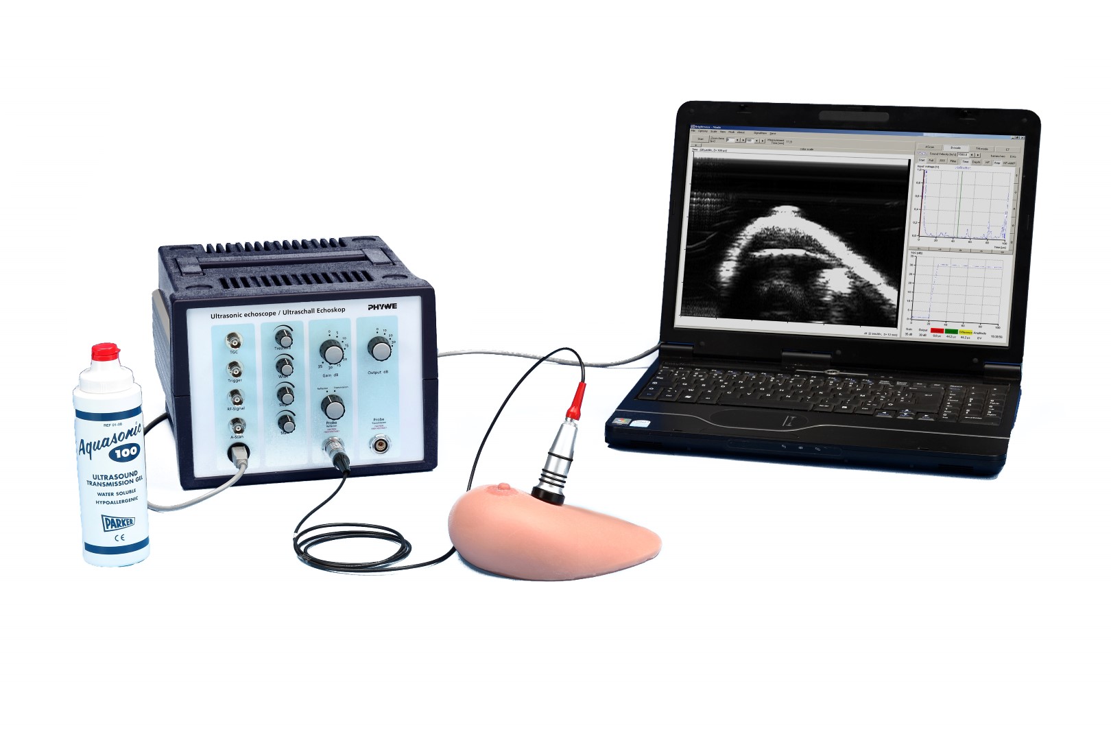
This experiment shows a typical application of ultrasound in medical diagnostics. A benign tumour on a realistic breast dummy is which has to be diagnosed, localized and measured with an ultrasound cross-section imaging method.
Benefits
- Ideal experiment for medical students in the preclinical phase: true-to-life breast cancer examinaton using a breast model
- The echoscope used in the experiment can also be used for other medically relevant experiments like A-scan, B-scan and ultrasound tomography
- Display of ultrasound image as for a diagnostic system
Tasks
- Examine the breast dummy and search for any pathological changes. Try to characterize them as accurately as possible (size, location, mobility, strength of the change).
- Produce an ultrasonic B-scan image of the breast dummy, especially in the regions of interest. Based on the ultrasound image, estimate the location and magnitude of the tumour.
Learning objectives
- Breast sonography
- Tumour size
- Benign Tumour
- Ultrasound imaging procedures
- Ultrasound echography
- A-mode
- B-mode
Device name
Article no.
Quantity
Name
File name
File size
File type
(en) Experiment guide
p5950300e .pdf
File size 0.52 Mb
pdf
Free shipping from 300,- €