Technical data Mitosis and Meiosis Set I, 6 selected microscope slidesArticle no: LIE-5150 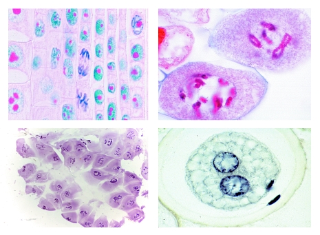
<!DOCTYPE html PUBLIC "-//W3C//DTD XHTML 1.0 Strict//EN"
"http://www.w3.org/TR/xhtml1/DTD/xhtml1-strict.dtd">
Function and Applications 5150 Mitosis and Meiosis Set I, - 6 selected Microscope SlidesMitosis, l.s. from Allium root tips showing all stages of plant mitosis carefully stained with iron-hematoxyline after Heidenhain - Mitotic stages in sec. through red bone marrow of mammal - Meiotic and mitotic stages in sec. of Salamandra testis. Many meiotic and mitotic stages can be observed - Lilium, anther t.s., microspore mother cells showing telophase of first and prophase of second (homeotypic) division - Giant chromosomes, smear from salivary gland of Chironomus, carefully fixed and stained * - Ascaris megalocephala embryology. Sec. of uteri showing maturation stages (meiosis). Polar bodies can be seen. The microslides are supplied in a slide box. |
PHYWE Systeme GmbH & Co. KG
Robert-Bosch-Breite 10 – 37079 Göttingen – Germany
www.phywe.com
Robert-Bosch-Breite 10 – 37079 Göttingen – Germany
www.phywe.com