Technical data Principles of Digital X-ray ImagingArticle no: P2550106 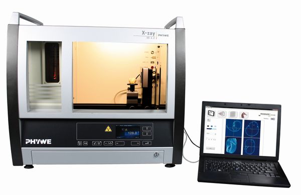
<!DOCTYPE html PUBLIC "-//W3C//DTD XHTML 1.0 Strict//EN"
"http://www.w3.org/TR/xhtml1/DTD/xhtml1-strict.dtd">
Principle With digital X-ray imaging, X-ray photons that interact with the detector are converted to a digital signal. This permits to record digital radiographies. With this experiment, the principles of digital detectors for X-ray imaging are laid out. Tasks
Learning objectives
Scope of delivery
| |||||||||
PHYWE Systeme GmbH & Co. KG
Robert-Bosch-Breite 10 – 37079 Göttingen – Germany
www.phywe.com
Robert-Bosch-Breite 10 – 37079 Göttingen – Germany
www.phywe.com