Technical data 3B MICROanatomy™ EyeArticle no: 3BS-1000260 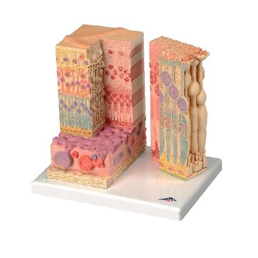
<!DOCTYPE html PUBLIC "-//W3C//DTD XHTML 1.0 Strict//EN"
"http://www.w3.org/TR/xhtml1/DTD/xhtml1-strict.dtd">
Function and Applications The MICROanatomy™ Eye model illustrates the microscopic anatomical structure of the retina with choroid and sclera. The left block-like, layered side of the eye model shows the complete structure of the retina including the supplying vascular layer and parts of the sclera from a light microscopic view.The right part of the eye model is a sectional enlargement. MICROanatomy™ Eye shows the microscopic structure of the photoreceptors and the cells of the pigmented layer.Left part of MICROanatomy™ Eye 850-times enlarged - right part 3800-times enlarged. You've never seen the human eye like this before! |
PHYWE Systeme GmbH & Co. KG
Robert-Bosch-Breite 10 – 37079 Göttingen – Germany
www.phywe.com
Robert-Bosch-Breite 10 – 37079 Göttingen – Germany
www.phywe.com