Function and Applications
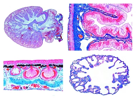
5900 Histology of the Frog (Rana sp.) - 20 Microscope SlidesFrog, lung t.s., simple sac-like respiratory organ - Frog, heart l.s. through the entire organ - Frog, blood smear, shows nucleated red corpuscles - Frog, spleen t.s., lymphatic tissue - Frog, tongue t.s., papillae, glands, muscles - Frog, esophagus t.s., shows ciliated epithelium - Frog, stomach t.s., glandular epithelium - Frog, small intestine t.s., folds of intestinal membrane, chyle - Frog, large intestine (colon) t.s., goblet cells - Frog, pancreas t.s., showing islets of Langerhans - Frog, liver t.s., showing liver parenchyma cells and bile ducts - Frog, kidney t.s., Malpighian corpuscles, renal vessels - Frog, urinary bladder t.s., smooth muscles, transitional epithelium - Frog, ovary t.s. shows follicle development, formation of yolk - Frog, testis t.s. showing spermatogenesis and mature spermatozoa - Frog, fallopian tube (Müllerian duct) t.s., glandular cells - Frog, interior brain t.s. showing nerve cells - Frog, spinal cord t.s., showing motor nerve cells - Frog, retina t.s., with rods and cones - Frog, skin t.s., skin glands, epidermis, pigment cells
The microslides are supplied in a slide box.